Abdominal Aortic Ultrasound
An ultrasound of the abdominal aorta is a non-invasive, painless test that uses high-frequency sound waves to image the “aorta,” the main blood vessel leading away from the heart. When the walls of the abdominal aorta become weak, they may balloon outward If the aorta reaches over 3 centimeters in diameter, it is then called an abdominal aortic aneurysm (AAA). As the aneurysm gets larger, the risk of rupture increases. Ultrasound imaging of the aorta is useful for measuring its size to screen for AAA.
Ablation
Catheter ablation (ab-LA-shun) is a medical procedure used to treat some arrhythmias (ah-RITH-me-ahs). An arrhythmia is a problem with the speed or rhythm of the heartbeat. During catheter ablation, a long, thin, flexible tube is put into a blood vessel in your arm, groin (upper thigh), or neck. This tube is called an ablation catheter. It’s then guided to your heart through the blood vessel. A special machine sends energy through the catheter to your heart. This energy finds and destroys small areas of heart tissue where abnormal heartbeats may cause an arrhythmia to start.
Ankle Brachial Index (ABI)
A simple way for your doctor to check how well your blood is flowing and check for peripheral artery disease, or PAD. The test compares the blood pressure at your ankle with the blood pressure at your arm.
Cardiac Catheterization
Cardiac catheterization is a medical procedure used to diagnose and treat certain heart conditions. A long, thin, flexible tube called a catheter is put into a blood vessel in your arm, groin (upper thigh), or neck and threaded to your heart. Through the catheter, doctors can perform diagnostic tests and treatments on your heart.
Cardiac CT
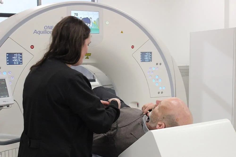
Cardiac computed tomography, or cardiac CT, is a painless test that uses an x-ray machine to take clear, detailed pictures of your heart. It’s a common test for showing problems of the heart. During a cardiac CT scan, the x-ray machine will move around your body in a circle and take a picture of each part of your heart. CT angiography is a method of visualizing the blood vessels in the heart, abdomen, and extremities.
CardioMEMS
CardioMEMS heart failure system is a miniaturized, wireless monitoring sensor that is implanted in the pulmonary artery during a minimally invasive procedure to directly measure PA pressure. The system allows patients to transmit pulmonary artery pressure data from their homes to their health care providers allowing for personalized and proactive management to reduce the likelihood of hospitalization.
Cardioversion
A medical procedure that restores a normal heart rhythm in people with certain types of abnormal heartbeats (arrhythmias). Cardioversion is usually done by sending electric shocks to your heart through electrodes placed on your chest.
Carotid Ultrasound
Carotid (ka-ROT-id) artery disease, which can lead to a stroke, is a condition in which a fatty material called plaque (plak) builds up inside the carotid arteries. You have two common carotid arteries—one on each side of your neck—that divide into internal and external carotid arteries. The internal carotid arteries supply oxygen-rich blood to your brain. The external carotid arteries supply oxygen-rich blood to your face, scalp, and neck. Carotid artery disease can be very serious because it can cause a stroke, or “brain attack.” A stroke occurs when blood flow to your brain is cut off. If blood flow is cut off for more than a few minutes, the cells in your brain start to die. This impairs the parts of the body that the brain cells control. A stroke can cause lasting brain damage, long-term disability, paralysis (an inability to move), or death.
Coronary Calcium Scan
In the U.S., despite improvements in survival rates, one in four men and one in three women still die within a year of a first heart attack. Moreover, a large number of these heart attack victims die within 28 days after the onset of symptoms and, in this group, about two-thirds die before ever reaching a hospital. This underscores not only a need for better recognition of the warning signs of a heart attack but also a tremendous need for more efforts targeting prevention. Given that the risk of cardiovascular disease events can be reduced dramatically with proper care, physicians face major challenges when it comes to figuring out just who is at risk of a major heart-related event. In 2002, the American College of Cardiology (ACC) held a conference on preventive cardiology that asked the question: How can we do better? One recommendation was that all adults undergo a simple, office-based assessment as the first step to identifying individuals at higher risk for coronary artery disease (CAD) and CAD events.
ECG
An electrocardiogram also called an EKG or ECG, is a simple test that detects and records the electrical activity of the heart. It is used to detect and locate the source of heart problems. An EKG shows how fast the heart is beating. It shows the heart’s rhythm (steady or irregular) and where in the body the heartbeat is being recorded. It also records the strength and timing of the electrical signals as they pass through each part of the heart.
Echocardiography
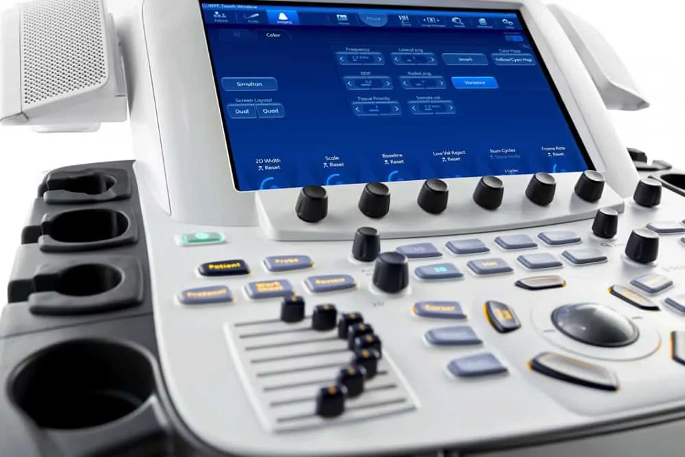
Echocardiography is a painless test that uses sound waves to create images of your heart. It provides your doctor with information about the size and shape of your heart and how well your heart’s chambers and valves are working.
Event Monitor
Holter and event monitors are medical devices that record the heart’s electrical activity. Doctors most often use these monitors to diagnose arrhythmias. Holter and event monitors also are used to detect silent myocardial ischemia. In this condition, not enough oxygen-rich blood reaches the heart muscle. “Silent” means that no symptoms occur.
Holter Monitor
Holter and event monitors are medical devices that record the heart’s electrical activity. Doctors most often use these monitors to diagnose arrhythmias. Holter and event monitors also are used to detect silent myocardial ischemia. In this condition, not enough oxygen-rich blood reaches the heart muscle. “Silent” means that no symptoms occur.
ICD and Pacemaker Check
Pacemaker and ICD Testing is done in our office on a daily basis. Both pacemakers and ICD’s (Implantable Cardioverter Defibrillators) implanted by our physicians at the hospital are checked with routine frequency in our Pacemaker/ ICD Clinic. It is important for a patient with a newly implanted device to keep the clinic appointment that is set when they leave the hospital. The patient may then be scheduled to come to the clinic for an appointment on a regular interval or may be scheduled to use an “at home” testing device. Important: If you have an ICD and it delivers a shock to you, please report it to our device nurses.
If you have frequent or several shocks in a brief time period, the recommendations for when an ICD shocks a patient are as follows:
- Single shock with no symptoms during business hours – call a device RN
- Single shock with symptoms – always go to the ER
- Multiple shocks – Go to the ER, regardless if we are open or not
- Any time the clinic is closed and a shock is delivered – go to the ER
ICD Implantation
An implantable cardioverter-defibrillator (ICD) is a small device that’s placed in your chest or abdomen. This device uses electrical pulses or shocks to help control life-threatening, irregular heartbeats, especially those that could lead the heart to suddenly stop beating (sudden cardiac arrest).
Implantable Loop Recorder
An implantable loop recorder, or ILR, is a heart recording device that is implanted (in a minor procedure) in the body underneath the chest skin. The machine works as an electrocardiogram (ECG), continuously picking up electrical signal from your heart. It has several uses. The most common ones include looking for causes of fainting, palpitations, very fast or slow heartbeats, and hidden rhythms that can cause strokes. All loop recorders come with a handheld activator that tells the loop recorder to save the signals collected over a certain time. We will make sure you know how to use the activator before you go home and when to make follow-up appointments. It is important to carry the activator with you at all times, so you can capture and record any episodes whenever you’re experiencing symptoms. You may keep your loop recorder for up to 2 or 3 years. When you no longer need it, you may have it removed in a similar procedure.
Mobile Cardiac Telemetry
Mobile cardiac telemetry (MCT) is a cardiac monitoring method that uses a small portable device to monitors a patient’s cardiac activity. It records the patient’s heartbeat as they run errands, exercise, and sleep.
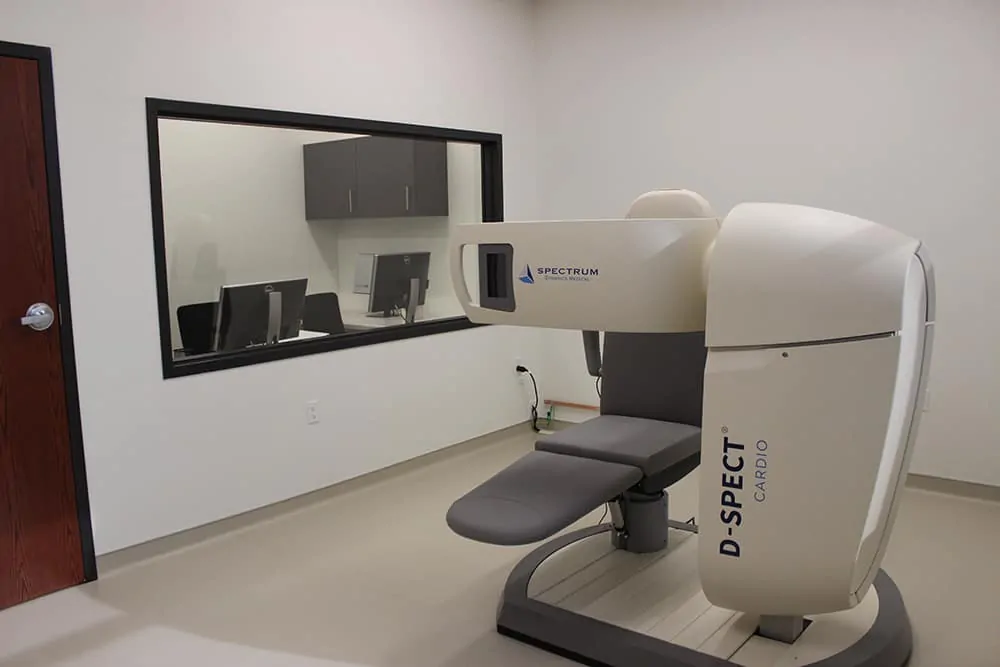
Nuclear Cardiac Testing
A nuclear heart scan is a type of medical test that allows your doctor to get important information about the health of your heart. During a nuclear heart scan, a safe, radioactive material called a tracer is injected through a vein into your bloodstream. The tracer then travels to your heart. The tracer releases energy, which special cameras outside of your body detect. The cameras use the energy to create pictures of different parts of your heart.
Pacemaker Implantation
A pacemaker is a small device that’s placed under the skin of your chest or abdomen to help control abnormal heart rhythms. This device uses electrical pulses to prompt the heart to beat at a normal rate. Pacemakers are used to treat heart rhythms that are too slow, fast, or irregular. These abnormal heart rhythms are called arrhythmias. Pacemakers can relieve some symptoms related to arrhythmias, such as fatigue (tiredness) and fainting. A pacemaker can help a person who has an abnormal heart rhythm resume a more active lifestyle.
Peripheral Intervention
Peripheral arterial disease (PAD) occurs when a fatty material called plaque builds up on the inside walls of the arteries that carry blood from the heart to the head, internal organs, and limbs. PAD is also known as atherosclerotic peripheral arterial disease.
Peripheral Vascular CT Angiography
Computed tomography angiography or CTA is a painless test that uses an x-ray machine to take 3-dimentionsional pictures of the arteries in peripheral vasculature of the body. This test is excellent for detecting and giving more detail to blockage of carotid arteries, kidney arteries, or pelvic and leg arteries.
Peripheral Vascular Ultrasound
An ultrasound is a non-invasive, painless test that uses high-frequency sound waves to image the arteries in the legs. These tests can detect deep arterial leg blockage that can lead to leg pain or eventually amputations in those patients at risk.
Rhythm Strip
This is a printed study of a patient’s EKG, which may indicate normal or abnormal conditions. Abnormal rhythms are called arrhythmia or sometimes, dysrhythmia. Arrhythmia is an abnormally slow or fast heart rate or an irregular cardiac rhythm. The EKG waveform has several pieces for each heartbeat.
Stress Echocardiography
An echocardiogram is a painless, harmless test that uses high-frequency sound waves (ultrasound) to examine the heart’s anatomy and function. A treadmill stress test evaluates your heart’s response to physical activity through the monitoring of your heart rate, blood pressure, and electrocardiograms while you exercise on a treadmill. When the two tests are combined, an assessment can be made of the status of your heart at rest, as well as during and immediately following stress. This can provide your doctor with information regarding whether or not you have significant blockages in your heart arteries. Other information obtained includes an assessment of the pumping function of your heart and the status of your heart valves.
Stress Testing
Stress testing provides your doctor with information about how your heart works during physical stress. Some heart problems are easier to diagnose when your heart is working hard and beating fast. During a stress test, you exercise (walk or run on a treadmill or pedal a bicycle) or are given a medicine to make your heart work harder while heart tests are performed.
TAVR
Transcatheter aortic valve replacement (TAVR) is a minimally invasive procedure to replace a narrowed aortic valve that fails to open properly (aortic valve stenosis). Transcatheter aortic valve replacement is sometimes called transcatheter aortic valve implantation (TAVI).
Tilt Table Testing
An arrhythmia (ah-RITH-me-ah) is a problem with the speed or rhythm of the heartbeat. During an arrhythmia, the heart can beat too fast, too slow, or with an irregular rhythm.
Transesophageal Echocardiogram
Echocardiography is a painless test that uses sound waves to create images of your heart. It provides your doctor with information about the size and shape of your heart and how well your heart’s chambers and valves are working.
Vein Ablation Procedure
An image-guided, minimally invasive treatment. It uses radiofrequency or laser energy to cauterize (burn) and close the abnormal veins that lead to varicose veins.
Vein Thrombosis and Obstruction Intervention
Sometimes deep vein thrombosis and obstruction of the larger veins in the pelvis are treatable with procedures to dissolve the clot and sometimes place large stents. These procedures are generally performed in the hospital setting. This disease entity is known as May-Thurner Syndrome.
Venous CT Angiography
Computed tomography angiography or CTA is a painless test that uses an x-ray machine to take 3-dimentionsional pictures of the veins in the abdomen, pelvis, and legs. This test is excellent for detecting obstruction in large pelvic veins (May-Thurner Syndrome) and other complex venous problems.
Venous Ultrasound
This ultrasound is a non-invasive, painless test that uses high-frequency sound waves to image the veins in the legs. These tests can detect deep vein thrombosis (DVT) and vein valve leakage (venous reflux).
Accreditations

Laboratory Services
Heart and Vascular Institute of Wisconsin offers a full-service laboratory department.

